 |
 |
||||||||||
| EXAFS | ||||||||||||||||||||||||||
Zr K-EDGE EXAFS STUDY OF PZT THIN FILM FORMATION FROM SOLS
Related publications: I. Arčon, B. Malič,
M. Kosec, A. Kodre; Zr K-Edge EXAFS Study of PZT Thin Film Formation From Sols,
Physica Scripta. Vol, T115, (2005), 448-449
Abstract
We examined the local environment of zirconium atoms in the Pb(Zr![]() Ti
Ti![]() )O
)O![]() (PZT) sol, prepared from lead acetate and transition metal n-propoxides in 2-methoxyethanol, and in the as-pyrolyzed amorphous film deposited on (0001) sapphire. The immediate neighborhood of Zr atoms changes markedly upon transition from the sol to the amorphous film. Clustering of Zr species in the process of film formation in the form of Zr oxide nanoparticles is indicated.
(PZT) sol, prepared from lead acetate and transition metal n-propoxides in 2-methoxyethanol, and in the as-pyrolyzed amorphous film deposited on (0001) sapphire. The immediate neighborhood of Zr atoms changes markedly upon transition from the sol to the amorphous film. Clustering of Zr species in the process of film formation in the form of Zr oxide nanoparticles is indicated.
Introduction
Ferroelectric thin films have been widely studied for a range of applications such as memories, capacitors, sensors or microelectromechanical devices [1,2]. In comparison to solid-state synthesis, alkoxide based sol-gel processing of multicomponent materials is recognized to yield more homogeneous products at lower processing temperatures, due to the formation of heterometallic bonding already in solution. In chemical solution deposition (CSD) of thin films the main processing steps include the synthesis of a heterometallic precursor, typically in a nonaqueous medium, the deposition of the film, and the processes occurring upon thermal treatment: drying, consolidation and crystallization of the target ferroelectric phase [3]. Understanding the structural transitions in the process of film formation from the sol would allow a better tailoring of the properties of the final product.
In 2-methoxyethanol-based processing of Pb(Zr![]() Ti
Ti![]() )O
)O![]() (Zr-rich PZT) thin films the inorganic network arises in the reaction between transition metal alkoxides and lead acetate. The thermodynamically stable perovskite film on sapphire (0001) substrate crystallizes at
(Zr-rich PZT) thin films the inorganic network arises in the reaction between transition metal alkoxides and lead acetate. The thermodynamically stable perovskite film on sapphire (0001) substrate crystallizes at
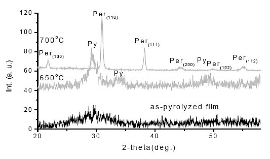
700![]() from the amorphous phase through the transitory pyrochlore-type phase [4] as generally observed for CSD PZT thin films [3]. It is known that the crystallization temperature of the perovskite phase is higher for Zr-rich PZT thin films than for Ti-rich thin films. Furthermore, the intermediate nanocrystalline pyrochlore-type (or fluorite) phase exists in a broader temperature range in Zr-rich PZT films [5,6]. So far there are no reports on the structure of the amorphous phase of the PZT films, even though there are indications that it crucially influences the perovskite crystallization temperature in the film.
from the amorphous phase through the transitory pyrochlore-type phase [4] as generally observed for CSD PZT thin films [3]. It is known that the crystallization temperature of the perovskite phase is higher for Zr-rich PZT thin films than for Ti-rich thin films. Furthermore, the intermediate nanocrystalline pyrochlore-type (or fluorite) phase exists in a broader temperature range in Zr-rich PZT films [5,6]. So far there are no reports on the structure of the amorphous phase of the PZT films, even though there are indications that it crucially influences the perovskite crystallization temperature in the film.
In this work Zr K-edge EXAFS of the amorphous Pb(Zr![]() Ti
Ti![]() )O
)O![]() thin film pyrolyzed at 350°C and its precursor prepared by the 2-methoxyethanol route is studied to provide insight into the structural changes in the process of the formation of the amorphous Zr-rich PZT film from the sol.
thin film pyrolyzed at 350°C and its precursor prepared by the 2-methoxyethanol route is studied to provide insight into the structural changes in the process of the formation of the amorphous Zr-rich PZT film from the sol.
Experimental
PZT sol, corresponding to the target composition of PbZr![]() Ti
Ti![]() O
O![]() with 10 mol% PbO excess, was prepared in a 100 ml batch by diluting zirconium and titanium n-propoxides in 2-methoxyethanol and by adding anhydrous lead acetate in the required quantity. The metal contents of all compounds were determined gravimetrically. The reaction mixture was heated to approximately 60°C to get a clear solution. After 2 hours of refluxing and distillation of the ester formed during the reaction a stable 0.5 M sol was obtained. All manipulations were performed in dry nitrogen atmosphere. PZT thin film was deposited on sapphire substrate (0001) by spin-coating and pyrolyzed at 350°C for 1 minute. The film thickness was about 200 nm. X-ray diffraction showed that the film was amorphous.
with 10 mol% PbO excess, was prepared in a 100 ml batch by diluting zirconium and titanium n-propoxides in 2-methoxyethanol and by adding anhydrous lead acetate in the required quantity. The metal contents of all compounds were determined gravimetrically. The reaction mixture was heated to approximately 60°C to get a clear solution. After 2 hours of refluxing and distillation of the ester formed during the reaction a stable 0.5 M sol was obtained. All manipulations were performed in dry nitrogen atmosphere. PZT thin film was deposited on sapphire substrate (0001) by spin-coating and pyrolyzed at 350°C for 1 minute. The film thickness was about 200 nm. X-ray diffraction showed that the film was amorphous.
Zr K-edge EXAFS spectra of the PZT sol and thin film were recorded at the X1 station in HASYLAB at DESY (Hamburg, Germany). A Si(311) double-crystal monochromator was used with 3 eV resolution at 18 keV. Harmonics were effectively eliminated by detuning the monochromator crystal using a stabilization feedback control.
Absorption spectrum of the PZT sol was measured in a standard transmission mode. The sol, concentrated to approximately 1 M solution in order to increase signal-to-noise ratio, was inserted in a liquid absorption cell with 0.5 mm lucite windows. Sample thickness of about 1mm was chosen to obtain total absorption thickness of about 2 above Zr K-edge. Empty absorption cell served for a reference spectrum measured in identical conditions. The intensity of the incident and transmitted beam was measured with Ar filled ionization cells. The standard stepping progression within [-250 eV, 1000 eV] interval relative to the Zr K-edge was adopted with an integration time of 1s/step.
Zr EXAFS spectrum of the PZT film was measured in a fluorescence detection technique using a 4-channel Ge fluorescence detector. The absorption spectra were obtained as the ratio of the fluorescence detector signal and the signal of the incident photon beam from the ionization cell filled with argon at ambient pressure. The same stepping progression was adopted as in the case of the sol with an integration time of 4s/step. To improve signal to noise ratio, eighteen consecutive runs were superimposed.
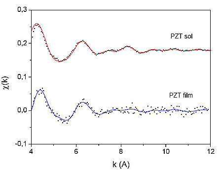
Figure 1: Zr K-edge EXAFS spectra of PZT sol and amorphous thin film on (0001) sapphire: (dots) - experiment, (solid line) - EXAFS model
Zirconium EXAFS spectra of PZT sol and thin film were analyzed by the University of Washington analysis programs using FEFF6 code for ab initio calculation of scattering paths [7,8]. The comparison of both spectra (Fig. 1) shows that the signal-to-noise ratio of the fluorescence EXAFS spectrum measured on the film is about an order of magnitude lower, in spite of the much longer detection time. Nevertheless, the quantitative analysis reveals that the local neighborhood of Zr atoms in the film is different from that observed in the sol. The difference can be clearly seen in Fig. 2, where the k![]() -weighted Fourier transforms of the EXAFS spectra are shown.
-weighted Fourier transforms of the EXAFS spectra are shown.
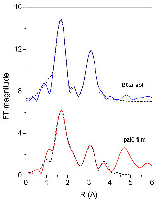
Figure 2: The k![]() weighted Fourier transforms of zirconium EXAFS spectra of PZT sol and amorphous thin film on (0001) sapphire, calculated in the k range of 4 - 11
weighted Fourier transforms of zirconium EXAFS spectra of PZT sol and amorphous thin film on (0001) sapphire, calculated in the k range of 4 - 11![]() : (Solid line)- experiment, (dashed line) - EXAFS model.
: (Solid line)- experiment, (dashed line) - EXAFS model.
Two shells of neighbors are discerned in the local neighborhood of Zr atoms in the sol. The fit of the EXAFS spectrum in the R range of 1.3![]() to 3.8
to 3.8![]() shows that the immediate neighborhood of Zr atoms is populated with about seven O atoms at two slightly different distances 2.11
shows that the immediate neighborhood of Zr atoms is populated with about seven O atoms at two slightly different distances 2.11![]() and 2.23
and 2.23![]() , while the wide second shell of neighbors consists of zirconium atoms at 3.47
, while the wide second shell of neighbors consists of zirconium atoms at 3.47![]() and carbon atoms at about 3.3
and carbon atoms at about 3.3![]() and 3.7
and 3.7![]() , in agreement with our previous study of PZT sols [4]. Carbon atoms belong to the functional groups of the starting compounds and the solvent. The number of Zr neighbors points to clustering of zirconium species.
, in agreement with our previous study of PZT sols [4]. Carbon atoms belong to the functional groups of the starting compounds and the solvent. The number of Zr neighbors points to clustering of zirconium species.
The local structure around Zr formed in the sol is not retained in the amorphous film. The fit of the EXAFS spectrum of the film (in the same R range of 1.3![]() to 3.8
to 3.8![]() ) shows that the distribution of oxygen atoms around Zr is significantly altered: there are about four oxygens at 2.14
) shows that the distribution of oxygen atoms around Zr is significantly altered: there are about four oxygens at 2.14![]() and about three at much larger distance of 2.67
and about three at much larger distance of 2.67![]() . In the second coordination shell Zr and O atoms are found, but no C atoms. The complete list of structure parameters is given in Table 1. Relatively high uncertainties in the number of neighbors in individual shells are due to the high correlation of the shell parameters N and
. In the second coordination shell Zr and O atoms are found, but no C atoms. The complete list of structure parameters is given in Table 1. Relatively high uncertainties in the number of neighbors in individual shells are due to the high correlation of the shell parameters N and ![]() .
.
Zr |
N |
R (A) |
s2 (A2) |
PZT sol |
|||
O |
3.8(5) |
2.11(4) |
0.002(1) |
O |
3.2(5) |
2.23(4) |
0.002(1) |
C |
3(1) |
3.29(1) |
0.003(1) |
Zr |
2(1) |
3.47(2) |
0.006(2) |
C |
5(2) |
3.63(5) |
0.003(1) |
PZT film |
|||
O |
4.0(7) |
2.14(4) |
0.003(1) |
O |
2.8(7) |
2.67(4) |
0.002(1) |
Zr |
3(1) |
3.43(2) |
0.006(2) |
O |
6(1) |
4.19(5) |
0.007(3) |
Table 1: Parameters of the nearest coordination shells around zirconium atom in PZT sol and amorphous thin film: atomic species, average number N, distance R and Debye-Waller factor ![]() . Uncertainty of the last digit is given in parentheses.
. Uncertainty of the last digit is given in parentheses.
We attribute the difference between the amorphous film and the sol to the partial decomposition of organic functional groups upon heating to 350![]() [3]. Namely, the local structure around Zr atoms in the film is similar to Zr neighborhood in the tetragonal ZrO
[3]. Namely, the local structure around Zr atoms in the film is similar to Zr neighborhood in the tetragonal ZrO![]() [9]. Most probably nano-clusters of zirconium oxide are forming within the film.
[9]. Most probably nano-clusters of zirconium oxide are forming within the film.
It is conceivable that the segregation of Zr species in the film - possibly as zirconia nanoparticles within the amorphous matrix - hinders the crystallization of the perovskite phase by the formation of the transient pyrochlore-type phase, so that the crystallization of the perovskite phase is shifted to higher temperature.
Acknowlwdgmwnt
Support by the Ministry of Science and Technology of the Republic of Slovenia within the National Research Program, by Internationales Buero BMBF (Germany), and by the IHP-Contract HPRI-CT-1999-00040 of the European Commision is acknowledged. L. Troeger and N. Haack of HASYLAB provided expert advice on beamline operation.
- Inoue N., IEEE Trans. On Electron Devices, 49, 1572 (2002).
- Muralt P., Micromech. Microeng. 10, 136 (2000).
- Tuttle B. A., Schwarz R. W., Mater. Res. Soc. Bull. 21, 49 (1996).
- Malič B., Kosec M., Arčon I., Kodre A., Hiboux S., and Muralt P., Integrated Ferroelectrics 20, 81 (2000)
- Huang Z., Zhang Q., Whatmore R. W., J. Appl. Phys. 85, 7355 (1999).
- Wilkinson A. P., Speck J. S., Ceetham A. K., Natarajan S., Chem. Mater. 6, 750 (1994).
- Stern, E. A., Newville, M., Ravel, B., Yacoby, Y. and Haskel, D., Physica B. 117, 208 (1995).
- Rehr, J. J., Albers, R.C. and Zabinsky, S.I., Phys. Rev. Lett. 69, 3397 (1992).
- Teufer, G., Acta Crystallographica 15, 1187 (1962).
|
|
|||||||||||||||||||||||||||||||||||||||||||||||||||||||||||
|
E-mail:iztok.arcon@p-ng.si Last change: 26-Jun-2006 |
|||||||||||||||||||||||||||||||||||||||||||||||||||||||||||